Exploring DNA Damage: A Hands-On Workshop on the Comet Assay
Dr. Kok Lun Pang
Comet Assay—a sensitive technique used to detect and quantify DNA damage at the level of individual cells.
This workshop is designed for students, researchers, and professionals in the fields of molecular biology, toxicology, environmental science, and biomedical research who wish to deepen their understanding of genomic integrity and genotoxic stress.
The Comet Assay, also known as single-cell gel electrophoresis (SCGE), is a widely used method for assessing DNA strand breaks and repair. This assay offers several advantages: it’s rapid, cost-effective, sensitive, and requires only a small number of cells.
Hands-on and Demonstration

Investigating Toxicity in Early Development: A Practical Workshop on Zebrafish (Danio rerio) Embryo Assays
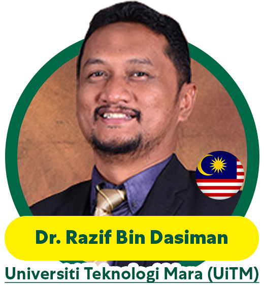
Dr. Razif Bin Dasiman
This hands-on workshop offers an in-depth introduction to the use of zebrafish (Danio rerio) embryos as a model system for assessing toxic effects of environmental and chemical exposures.
This workshop is designed for students, early-career researchers, and professionals in toxicology, pharmacology, environmental sciences, and developmental biology, the course combines foundational theory with practical training in embryo handling, exposure experiments, and endpoint analysis.
Zebrafish are a widely used vertebrate model in biomedical and environmental research due to their genetic similarity to humans, transparent embryos, rapid development, and high reproductive rate. The zebrafish embryo toxicity test (ZFET) has become a key tool in ecotoxicology and chemical safety assessment, offering a cost-effective, ethical alternative to mammalian models.
Toxic substances can disrupt normal development, leading to observable morphological and behavioral abnormalities in embryos. By studying these effects, researchers can evaluate the teratogenic, cytotoxic, or neurotoxic potential of compounds.s.

Histopathological Techniques in Zebrafish: Detecting Tissue Damage
Dr Fatin Nadzirah Binti Zakaria
This hands-on workshop introduces participants to histopathological staining methods for assessing inflammation, tissue integrity, and organ-specific damage in zebrafish (Danio rerio).
Designed for researchers and students in pathology, toxicology, aquatic biology, and biomedical sciences, the workshop blends core theory with practical laboratory skills in tissue preparation, staining, and microscopic evaluation.
Histopathology—the microscopic study of diseased tissue—is a cornerstone of toxicological and biomedical research. In zebrafish, which share conserved organ systems with humans, histopathology is a powerful tool for detecting subtle and chronic effects of chemical exposure, infections, or genetic mutations that are not visible through gross morphology alone.
Through proper tissue fixation, sectioning, and staining, researchers can detect cellular inflammation, necrosis, fibrosis, hyperplasia, and organ-specific damage in tissues such as liver, gills, intestines, kidney, and skin.
Hands-on and Demonstration
